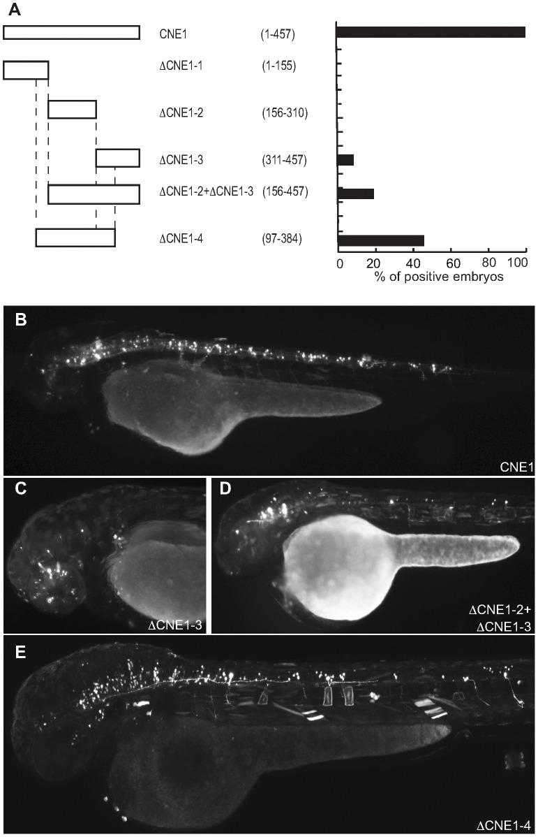
Figure 5. Deletion analysis of Meis_ciCNE1 construct.
(A) Scheme and quantification of the deletion constructs injected into zebrafish embryos and analysed for GFP expression at 48 hpf. The numbers in parentheses indicate the length of each construct. (B) Embryo injected with the full length CNE1 show GFP expression in the brain and in spinal cord motor- and interneurons. (C) Injection of DCNE1-3 drives GFP expression only in a few neurons in the head. (D) In the embryos injected with DCNE1-2+DCNE1-3 construct few motor- and interneurons are detected along the spinal cord. (E) In embryos injected with the construct DCNE1-4 GFP expression shows a pattern similar to full length, labelling both spinal cord interneurons and motor neurons as well as cells in the brain.

