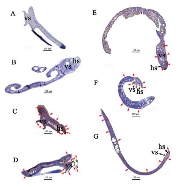
Fig. 3. Immunohistochemical staining for human telomerase reverse transcriptase in Schistosoma japonicum schistosomules exposed to VSVG-pseudotyped pBABE-puro- hTERT virions.
(A, B) Control, non-virion exposed schistosomules (showing head sucker, ventral sucker and posterior parts of the worms): absence of reaction for hTERT in sub-tegumental regions. (C, D, E, F, G) Schistosomules exposed to virions: intense staining for hTERT mainly located in sub-tegumental regions of the head sucker, ventral sucker and posterior tissues of the parasites as indicated by the arrows (red). Scale bar, 100 μm. hs, head (oral) sucker; vs, ventral sucker.

