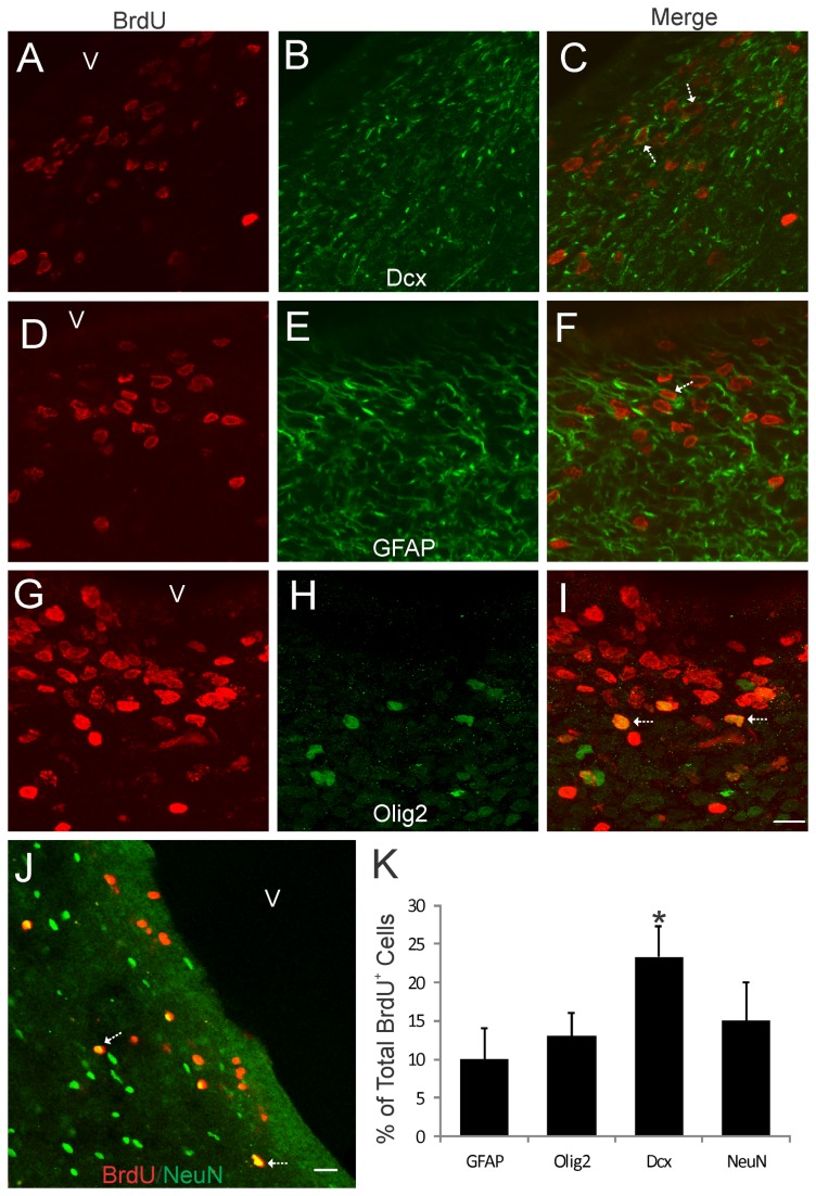
Figure 5. Bromodeoxyuridine (BrdU) cell proliferation tracking in piglet forebrain SVZ.
Confocal microscope images showing that subsets of BrdU+ cells in the forebrain SVZ can be immunophenotyped (arrows) as cells positive for the neuroblast marker doublecortin (A-C, Dcx), the astrocyte marker GFAP (D-F), the oligodendrocyte marker Olig2 (G-I), and the neuron marker NeuN (J). The lateral ventricle is identified (v). Scale bars: (in I, applies to A-I) = 12 µm; (in J) = 23 µm. K. Graph showing the proportions of the total BrdU+ cells that are either GFAP+ Olig2+, Dcx+, or NeuN+. Values are mean ± SD (n=4). Asterisk denotes Dcx significant difference (p < 0.05) compared to GFAP, Olig2, and NeuN.

