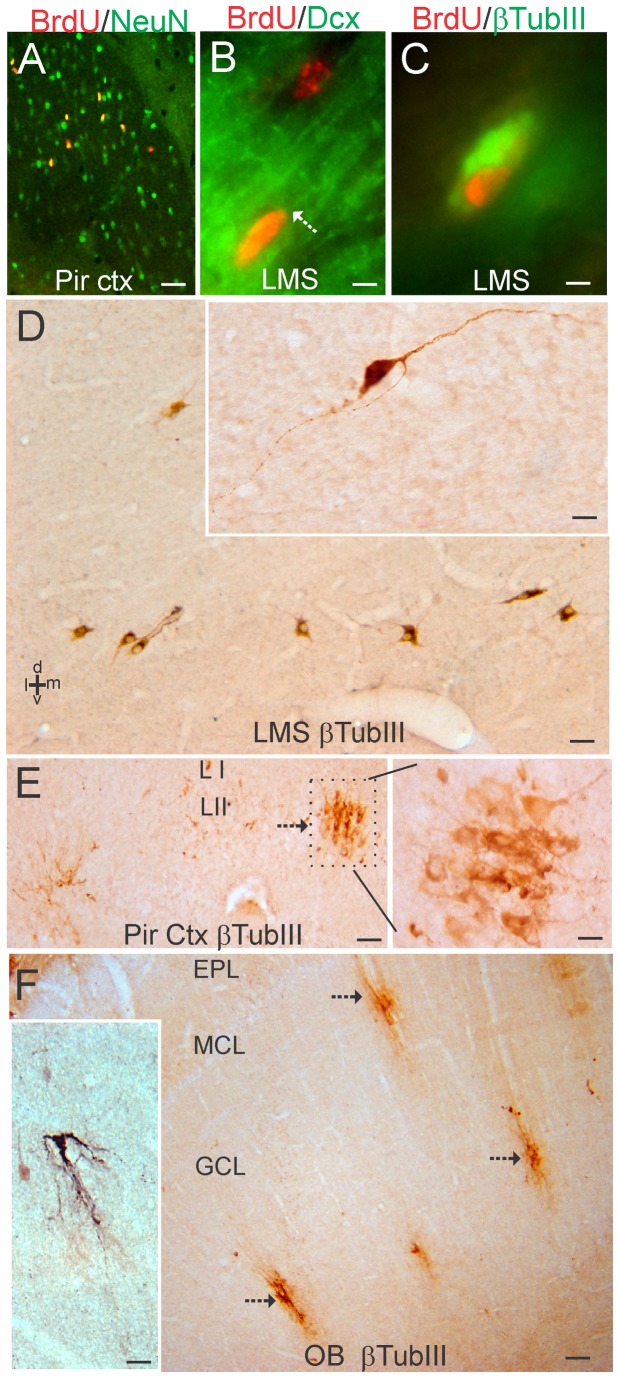
Figure 7. Characterization of the lateral migratory stream (LMS) of newly born neurons and identification of immature neurons in piglet forebrain.
A. Confocal image showing the colocalization of BrdU (red) and NeuN (green) in subsets of neurons in the piriform cortex. Single-labeled neurons are green, single-labeled BrdU+ nuclei are red, and double-labeled neurons are yellow. B. Immunofluorescence showing the colocalization of BrdU (red) and doublecortin (Dcx, green) in the LMS. One Brdu+ cell (top cell) does not express Dcx, but another Brdu+ cell (bottom cell) is Dcx+ as seen by the yellow around the nucleus and the trailing Dcx-labeled process (arrow). C. Immunofluorescence showing the colocalization of BrdU (red) and β-tubulin III (βTubIII, green) in the LMS. D. Immmunoperoxidase staining for β-tubulin III (βTubIII) revealed a column of βTubIII+ immature neurons (brown cells) below the anterior striatum within the LMS. Inset shows a βTubIII+ LMS neuron with a fusiform morphology and leading and trailing processes. E. In piriform cortex, βTubIII+ immature neurons were found in cellular islands in layer II (arrow). Boxed area is shown at right at higher magnification and rotated 90°. F. In OB, βTubIII+ immature neurons (arrows) were found in the granule cell layer (GCL). These cells possessed extensive local vertical-oriented arborizations (inset) consistent with an interneuron morphology [46]. EPL, external plexiform layer; MCL, mitral cell layer. Scale bars = 25 µm (A), 5 µm (B), 4 µm (C), 25 µm (D), 8 µm (D inset), 32 µm (E), 12.5 µm (E right), 100 µm (F), 25 µm (F inset).

