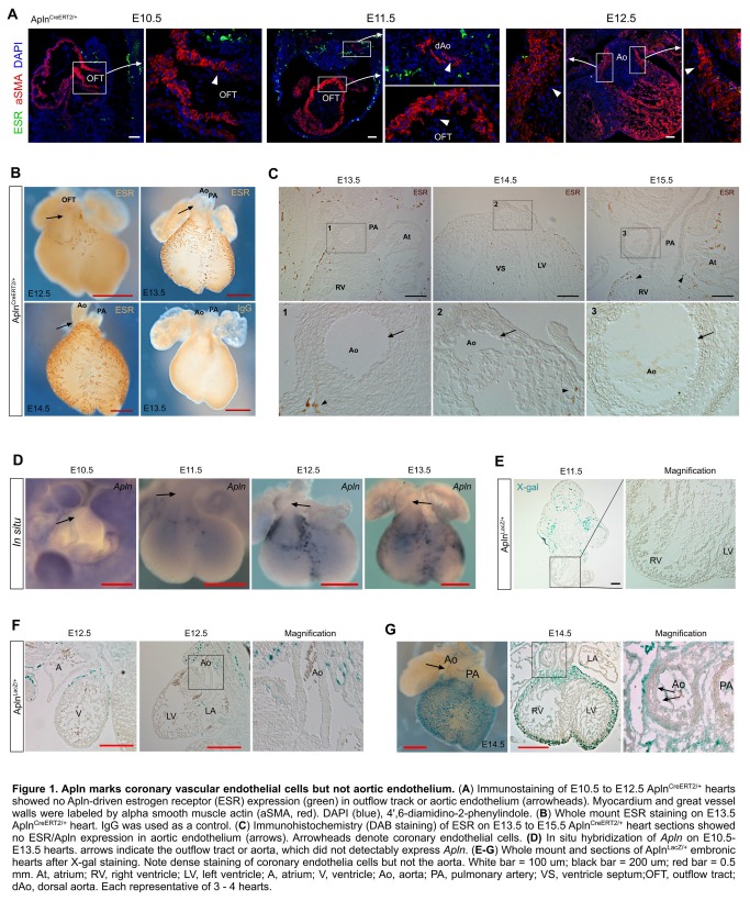
Figure 1. Apln marks coronary vascular endothelial cells but not aortic endothelium.
(A) Immunostaining of E10.5 to E12.5 AplnCreERT2/+ hearts showed no Apln-driven estrogen receptor (ESR) expression (green) in outflow track or aortic endothelium (arrowheads). Myocardium and great vessel walls were labeled by alpha smooth muscle actin (aSMA, red). DAPI (blue), 4',6-diamidino-2-phenylindole. (B) Whole mount ESR staining on E13.5 AplnCreERT2/+ heart. IgG was used as a control. (C) Immunohistochemistry (DAB staining) of ESR on E13.5 to E15.5 AplnCreERT2/+ heart sections showed no ESR/Apln expression in aortic endothelium (arrows). Arrowheads denote coronary endothelial cells. (D) In situ hybridization of Apln on E10.5-E13.5 hearts. Arrows indicate the outflow tract or aorta, which did not detectably express Apln. (E-G) Whole mount and sections of AplnLacZ/+ embryonic hearts after X-gal staining. Note dense staining of coronary endothelia cells but not the aorta. Arrows indicate aorta endothelium that is negative for Apln expression. White bar = 100 um; black bar = 200 um; red bar = 0.5 mm. At, atrium; RV, right ventricle; LV, left ventricle; A, atrium; V, ventricle; Ao, aorta; PA, pulmonary artery; VS, ventricle septum;OFT, outflow tract; dAo, dorsal aorta. Each representative of 3 - 4 hearts.

