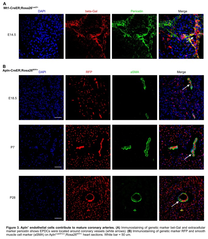
Figure 3. Apln+ endothelial cells contribute to mature coronary arteries.
(A) Immunostaining of genetic marker bet-Gal and extracellular marker periostin shows EPDCs were located around coronary vessels (white arrows). (B) Immunostaining of genetic marker RFP and smooth muscle cell marker (aSMA) on AplnCreERT2/+;Rosa26RFP/+ heart sections. White bar = 50 um.

