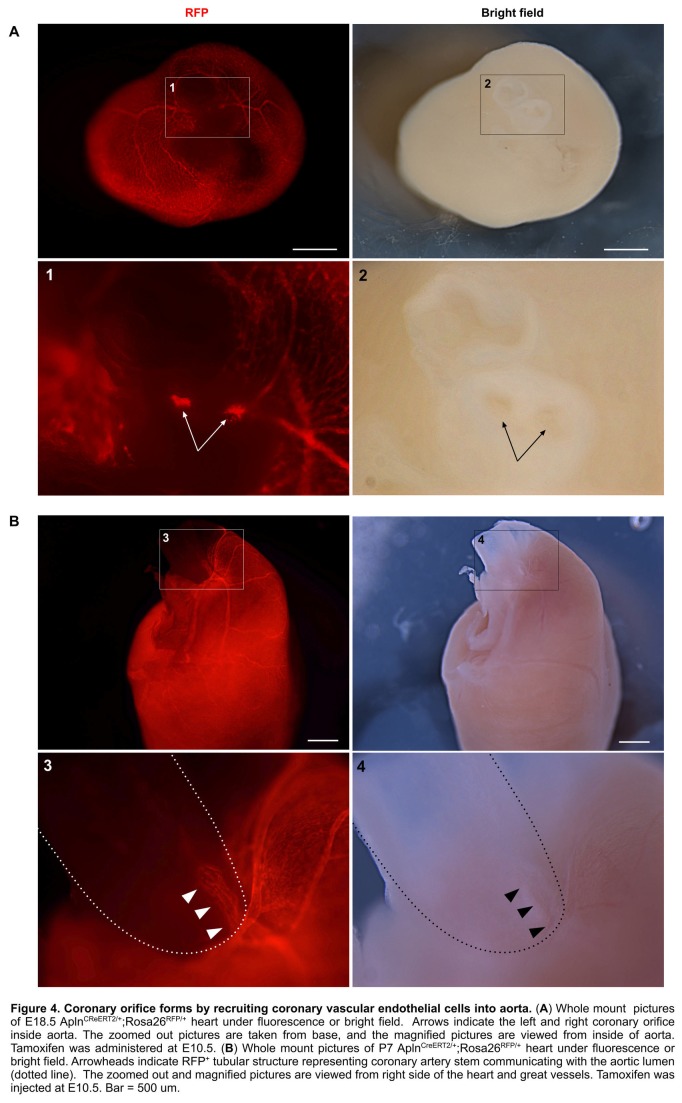
Figure 4. Coronary orifice forms by recruiting coronary vascular endothelial cells into aorta.
(A) Whole mount pictures of E18.5 AplnCreERT2/+;Rosa26RFP/+ heart under fluorescence or bright field. Arrows indicate the left and right coronary orifice inside aorta. The zoomed out pictures are taken from base, and the magnified pictures are viewed from inside of aorta. Tamoxifen was administered at E10.5. (B) Whole mount pictures of P7 AplnCreERT2/+;Rosa26RFP/+ heart under fluorescence or bright field. Arrowheads indicate RFP+ tubular structure representing coronary artery stem communicating with the aortic lumen (dotted line). The zoomed out and magnified pictures are viewed from right side of the heart and great vessels. Tamoxifen was injected at E10.5. Bar = 500 um.

