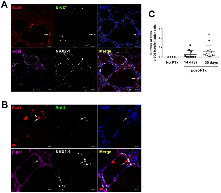
Figure 4. Immunofluorescence of BrdU, Sca1, NKX2-1, and β-gal in 35 and 120 days post-PTx thyroids.
(A) Immunofluorescence in 35 days post-PTx thyroid. Sca1(+);BrdU(+), but β-gal(−);NKX2-1(−) cell is located in an irregular shaped follicle (shown by an arrow). (B) Immunofluorescence in 120 days post-PTx thyroid. Sca1(+);BrdU(+) cell also expresses β-gal and NKX2-1 (shown by an arrow). Arrowhead in the Sca1 panel in B indicates cilia that are strongly positive for Sca1 due to the cell having these cilia is Sca1(+). Merged image is shown on the lower right. Scale bar: 20 µm. (C) Number of Sca1(+);β-gal(−) cells in 14 and 35 days post-PTx thyroids. The number shown is based on 1000 intrafollicular cells counted. N = 7–10.

