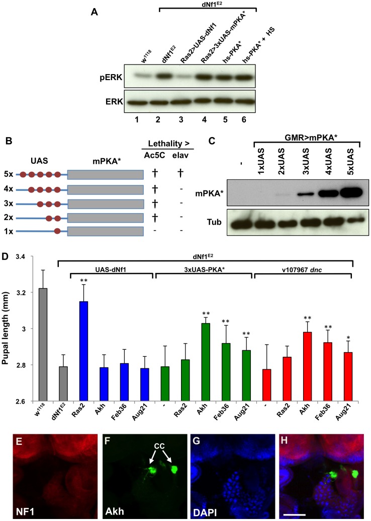
Figure 8. dNf1 systemic growth related RAS/ERK and cAMP/PKA signals appear functionally and topographically distinct.
(A) The elevated larval CNS pERK level of dNf1 mutants is reduced by neuronal expression of dNf1, but not by neuronal or heat-shock induced ubiquitous expression of PKA*. Western blot of pERK levels in larval CNS of the indicated genotypes. In lane 6, larvae received a daily 20 min 37°C heat shock throughout development, a protocol that suppresses the dNf1 growth defect [4]. (B) Structure of UAS-PKA* transgenes with 1 to 5 UAS elements. The lethality of these transgenes when driven with either Ac5C-Gal4 or elav-Gal4 is indicated by † whereas (−) indicates viable offspring. (C) Western blot of adult head lysates showing relative expression of GMR-Gal4-driven transgenic PKA*. Tubulin is used as a loading control. (D) Expression of PKA* or knockdown of dnc by shRNAi in the ring gland rescues the dNf1 pupal size defect. In contrast, UAS-dNf1 expression with the same ring gland drivers fails to restore systemic growth. (E–H) Expression pattern of Akh-Gal4 driving UAS-GFP, co-stained with DAPI and anti-dNF1. GFP expression in the corpora cardiaca (CC) is indicated. Scale bar = 50 µm. As previously noted [74], anti-dNf1 staining is strong in the CNS, whereas staining in the ring gland is close to background.

