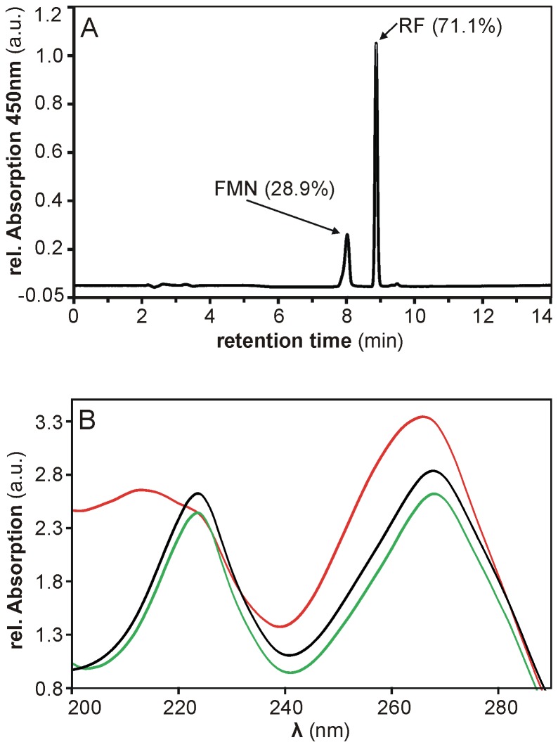
Figure 7. HPLC-based analysis of flavins isolated from B. subtilis cell lysate.
(A) By means of reverse-phase HPLC, flavins isolated from B. subtilis cell lysate using His-tagged Apo-YLOV as a molecular probe were identified as RF and FMN. The corresponding peaks are labeled by type of flavin and its relative amount. (B) Comparison of UV-Vis spectra measured at the respective peak maxima of FMN from B. subtilis (black line) and FMN (green line) and FAD (red line) from reference samples indicates that the faster eluting flavin species isolated from B. subtilis is indeed FMN.

