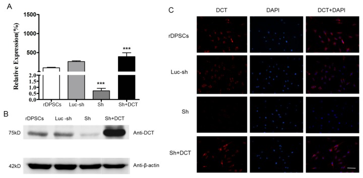
Figure 2. The silencing effect of siRNA on CaV1.2 channels and the rescue effect of DCT in CaV1.2 knockdown rDPSCs.
5 days after transfection, mRNA and protein expression of DCT were analyzed using real-time PCR and western blotting, respectively. The primer was designed at the end of C-terminus of CaV1.2 so that it can detect both the CaV1.2 mRNA and recombinant DCT mRNA. Sh cells: CaV1.2 knockdown cells. Luc-sh cells: luc-shRNA lentiviral control cells. ShSh+DCT cells: DCT-rescued ShSh cells generated by transfecting CaV1.2 knockdown cells with recombinant DCT lentiviral vector.
(A): mRNA levels of DCT in CaV1.2 knockdown rDPSCs and DCT-rescued ShSh cells.
(B): expression of DCT in CaV1.2 knockdown rDPSCs and DCT- rescued ShSh cells.
(C): Immunofluorescence of rDPSCs grown 5 days in vitro. Anti- DCT staining is shown in red and nuclei are shown in blue (Scale bars 100μm).

