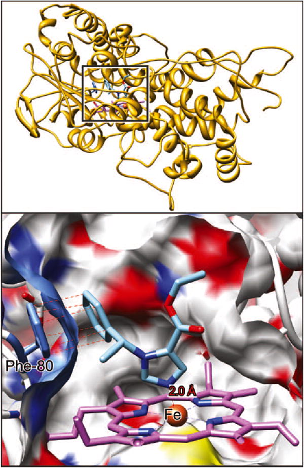
Figure 3.

Etomidate docked in the substrate-binding pocket of 11β-hydroxylase. A cross-section through the surface of the binding cavity is shown. The surface is colored according to the atoms behind it; white is carbon, blue nitrogen, red oxygen, and yellow sulfur. Etomidate is depicted in stick representation with blue nitrogens, red oxygens, and sky blue carbons. In the heme system, purple is carbon, blue is nitrogen, and then rust is heme. The heme iron atom is coordinated to Cys-400 below the ring and etomidate imidazole nitrogen above the ring. Top right insert from Figure 1 for reference.
