
Figure 5.
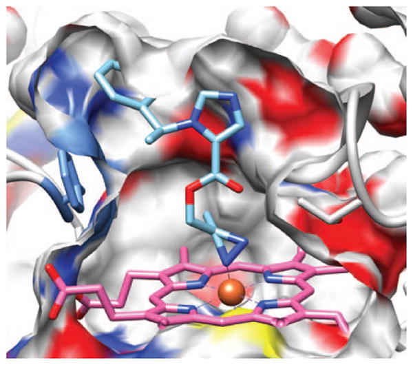
Representative pose showing azi-etomidate docked in the substrate-binding pocket of 11β-hydroxylase with its azi moiety interacting with the enzyme's heme iron. A cross-section through the surface of the binding cavity is shown. The surface is colored according to the atoms behind it; white is carbon, blue nitrogen, red oxygen and yellow sulfur. Azi-etomidate is depicted in stick representation with blue nitrogens, red oxygens, and sky blue carbons.