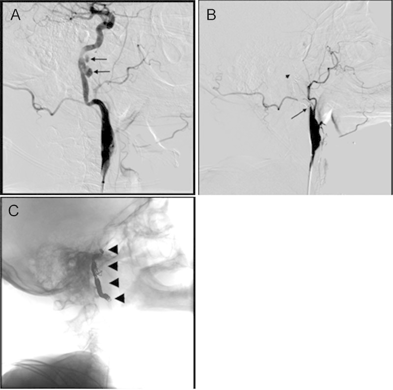
Fig. 2.

Angiography. (A) Pretreatment image shows a multilobulated pseudoaneurysm (arrows) of the cervicopetrous internal carotid artery (ICA). (B) Posttreatment image showing no filling of the pseudoaneurysm. Long arrow located at the proximal ICA. Short arrow demonstrates the occluded pseudoaneurysm. (C) Posttreatment image showing coil placement (arrowheads) from the petrous ICA to the proximal ICA.
