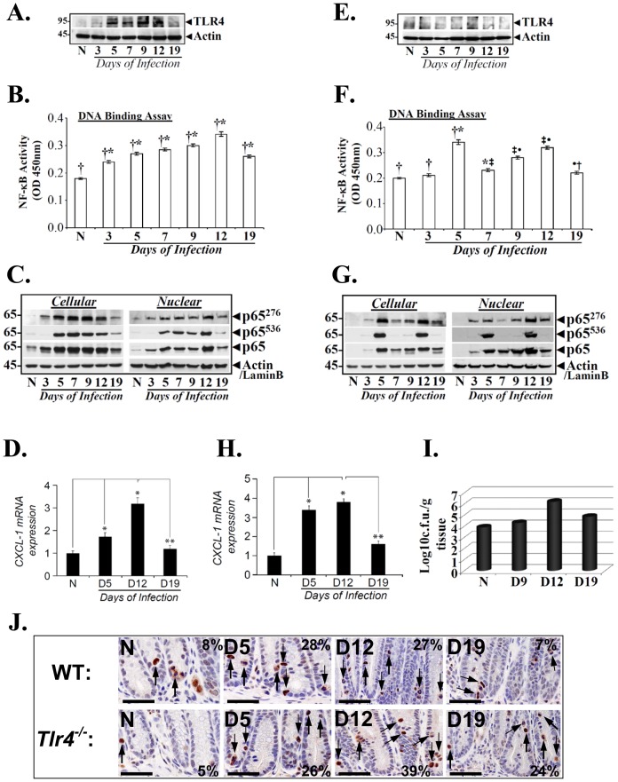
Figure 2. TLR4-dependent and independent mechanism of NF-κB activation during TMCH.
Relative levels of TLR4 in the cellular extracts prepared from the isolated crypts of uninfected normal (N) and CR infected (days 3–19) C57Bl/6 (A) and Tlr4−/− (E) mice, respectively. Actin was used as internal control. Measurement of NF-κB activity in the colonic crypts of C57Bl/6 and TLR4−/− mice. NFκB-p65 activity in the nuclear extracts prepared from the uninfected normal (N) and days 3–19 post infected C57Bl/6 (B) and Tlr4−/− (F) mice was examined by utilizing TransAM NF-κB p65 Chemi Transcriptional Factor assay kit from Active Motif. †*, p<0.05 vs. N; *‡, p<0.05 vs. day 5; ‡•, p<0.05 vs. day 7; •†, p<0.05 vs. day 12 (n = 3 independent experiments). Phosphorylation status of p65 subunit in the colonic crypts of C57Bl/6 and Tlr4−/− mice. Relative levels of phosphorylated (p65276/p65536) and total p65 subunit in the cellular and nuclear extracts prepared form the uninfected normal (N) and days 3–19 post infected C57Bl/6 (C) and Tlr4−/− (G) mice were determined by Western blotting with moiety-specific antibodies. Actin or LaminB were loading controls. CXCL-1/KC expression in the colonic crypts of C57Bl/6 and Tlr4−/− mice. Expression of CXCL-1/KC mRNA isolated from the crypts of uninfected normal (N) and days 3–19 post infected C57Bl/6 (D) and TLR4−/− (H) mice respectively, was measured as readout for NF-κB activity, via real-time RT-PCR. *, p<0.05 vs. N; **, p<0.05 vs. day 12, (n = 3 independent experiments). I. Bacterial counts correlate with NF-κB activation in Tlr4−/− mice. Distal colonic homogenates, prepared from the uninfected and CR-infected (days 9–19) Tlr4−/− mice were plated on McConkey agar and incubated at 37°C. Pink Citrobacter rodentium colonies were counted and plotted as log values (n = 3 independent experiments). J. Immunohistochemical staining of p65 subunit phosphorylated at Ser-276 in vivo. Paraffin embedded sections prepared from the distal colons of uninfected normal (N) and days 5, 12 and 19 post-infected wild type C57Bl/6 (WT) and Tlr4−/− mice were stained with antibody specific for NFκB-p65 phosphorylated at Ser-276 and were analyzed with light microscopy. Percentages represent percent cells positive for p65276 staining. (Scale bar = 75 µm; n = 3 independent experiments).

