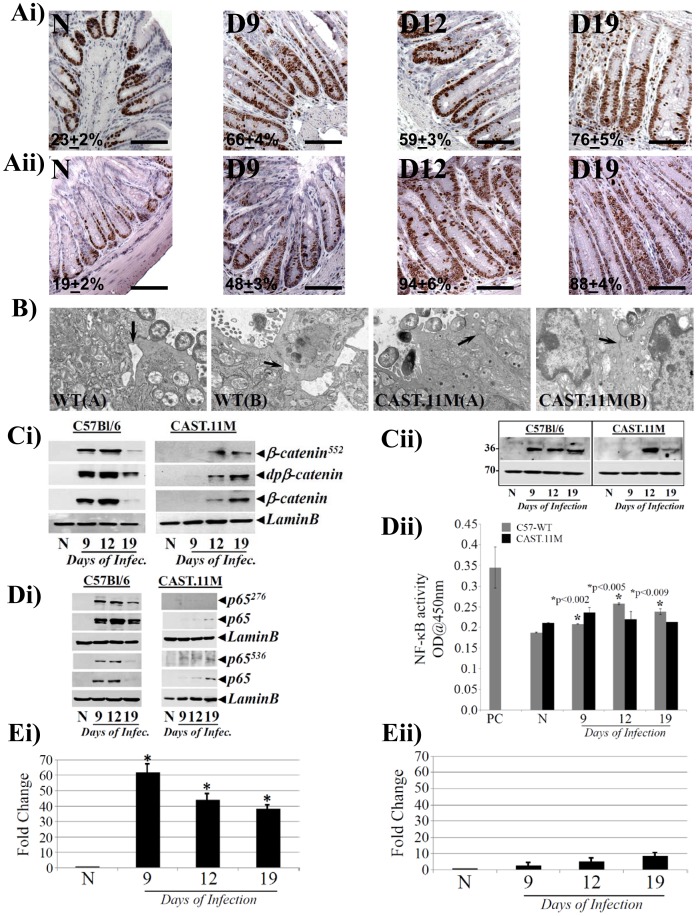
Figure 8. B6.CAST.11M mice exhibit a hyperplastic but a muted inflammatory response to CR infection.
A. Representative photomicrographs of paraffin embedded sections prepared from the distal colons of uninfected normal (N) and days 9–19 post-CR infected C57Bl/6 (upper panel) or CAST.11M (lower panel) mice and stained for Ki-67 (percentages represent percent cells positive for Ki-67). Scale bar: 50 µm; n = 3 independent experiments. B. Electron Microscopy. Distal colonic fragments from CR infected wild type C57Bl/6 (WT) or CAST.11M mice were subjected to transmission electron microscopy to detect disruption in tight junctions (arrows). Two representative images are shown for each group of mice. C. Relative levels of phosphorylated (β-Cat552), de-phosphorylated (dpβ-Catenin) and total β-catenin (Ci) and cyclinD1 (Cii) in the nuclear extracts prepared form the uninfected normal (N) and days 9, 12 and 19 post infected wild type C57Bl/6 and B6.CAST.11M mice, were determined by Western blotting with moiety-specific antibodies. LaminB was used as loading control. Di. Nuclear extracts prepared from the distal colonic crypts of groups of mice described in C were probed with antibodies for: p65276, p65536 and total p65, respectively. LaminB was used as loading control. Dii. Measurement of NF-κB activity. NF-κB-p65 activity in the nuclear extracts prepared from the groups of mice described in C was measured by utilizing TransAM NF-κB p65 Chemi Transcriptional Factor assay kit from Active Motif. E. CXCL-1/KC expression in the colonic crypts. Expression of CXCL-1/KC mRNA isolated from the distal colonic crypts of C57Bl/6 (Ei) or CAST.11M (Eii) mice was measured as readout for NF-κB activity, via real-time RT-PCR. *, p<0.05 vs. N; n = 3 independent experiments.

