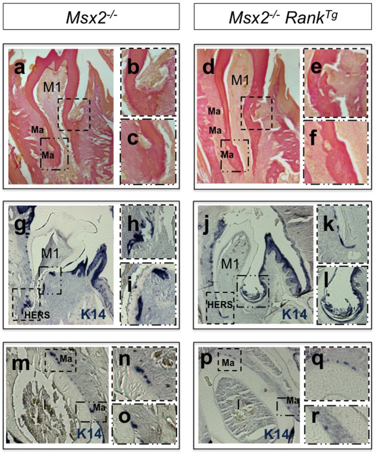
Figure 3. Effect of transgenic Rank on lower first molar and incisor root formation in Msx2 − /− mice.
Van Gieson histology staining (a–f) and keratin immunohistochemistry (g–r) were respectively performed on mandibular frontal sections of 3- and 2-week-old Msx2 −/− mice either overexpressing or not expressing transgenic Rank. At 3 weeks, Rank overexpression had induced a normalization in the size of most epithelial cell rests of Malassez (Ma) (a and c versus d and f), and at 2 weeks it had induced a better commitment of Hertwig epithelial root sheath (HERS) cells, specifically in the labial area (j and k versus g and h). Occasionally and independently of Rank overexpression, epithelial cyst–like structures were observed in the lingual area of Msx2 −/− mandibular first molars (j, i). Cytokeratin-14 immunolabelling revealed that these cyst-like structures were associated with abnormal continuity between dental and oral epithelia (j) and the presence of a periodontal pocket (square in j enlarged in l). Another defect observed at 3 weeks in the lingual root of Msx2 −/− mice, also independent of Rank overexpression, was a lacuna-like structure in the dentine facing the site of transition between crown and root epithelia (squares in a and d enlarged in b and e, respectively). In the incisor root equivalent, normalization of the size of the epithelial cell rests of Malassez (Ma) was observed in Msx2 −/− RankTg mice (squares in m versus p enlarged in n and o and q and r, respectively).

