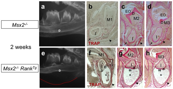
Figure 4. Effect of Rank overexpression on lower incisors of Msx2 − /− mice.
Mandibular microradiographs (a, e) and TRAP activity assays (b, f) or van Gieson histology staining (c, d, g, h) of mandibular frontal sections were performed to characterize the effect of Rank overexpression on the lower incisors of 2-week-old Msx2 −/− mice. Substantial enlargement of the area between the basal bone and the dentin was observed in Msx2 −/− RankTg mice (e) compared to Msx2 −/− mice not expressing RankTg (a). This enlarged area, which corresponds to the incisor epithelial compartment, was associated with abnormal curvature in both basal bone (red dotted line) and dentin (D). Mandibular frontal sections through the first (M1), second (M2), and third (M3) molar planes revealed that the enlargement corresponds to an epithelial hypertrophy with the presence of an internal necrosis-like area (asterisks in f–h). In Msx2 −/− incisors, no hypertrophy of the epithelium was visible, but this tissue was disorganized and lacked the ameloblastic palisade structure (arrows in b–d). There was also a substantial increase in the number of osteoclasts around the incisors of Msx2 −/− RankTg mice (f) compared to Msx2 −/− mice not expressing RankTg (b). Moreover, the thickness of the mandibular basal bone in the Msx2 −/− RankTg mutants appeared highly reduced compared to Msx2 −/− mice not expressing RankTg (arrowheads in f–h versus b–d).

