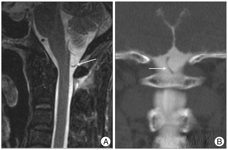
Fig. 1.
A : Sagittal, fast-spin echo T2-weighted magnetic resonance images of the occipito-cervical junction showing the anomalous course of the left posterior inferior cerebellar artery (arrow). B : Coronal, reformatted computed tomography-myelogram images of the occipito-cervical junction showing the tortuous and caudal course of the posterior inferior cerebellar artery as it closes the C1 lamina (arrow).

