
Fig. 3.
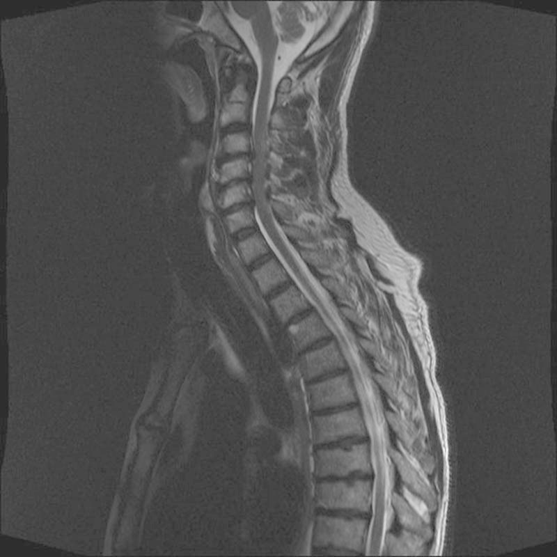
Sagittal T2-weighted magnetic resonance imaging of the cervical spine, which demonstrated moderately severe spinal stenosis at the C3–C4, C4–C5, and C5–C6 levels, with less severe spinal stenosis at the C5–C6 level. There was also increased signal in the anterior vertebral body of T3, which were thought to represent small hemangiomas