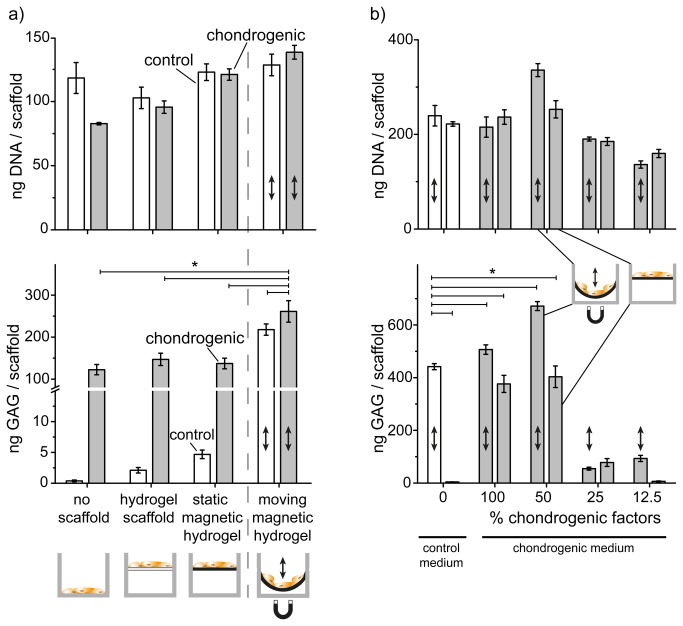
Figure 3. Mechanical stimulation induced chondrogenesis.
a) Cell numbers (DNA amount per scaffold) confirmed good cell expansion and growth. Below is the glycosaminoglycan (GAG) deposition per scaffold over a period of 5 weeks. Control medium (white bars) and chondrogenic medium (grey bars) were applied on cells seeded into either tissue culture plate (no scaffold), hydrogel scaffold (no nanomagnets, i.e. no movement is possible) or magnetic hydrogel. Mechanical stimulation (arrow) triggered higher GAG deposition. b) Comparable DNA amount indicated good cell growth for cells seeded into magnetic hydrogels with both medium types and no negative effects from mechanical stimulation. GAG deposition using diluted chondrogenic (grey) versus control medium (white bars). Mechanically stimulated hMSC in control medium showed comparable GAG deposition as in standard chondrogenic medium under magnetic actuation (indicated by ↕). * p < 0.01 cells cultured with control medium under mechanical stimulation versus non stimulated and mechanically stimulated hydrogel using both cell culture media.

