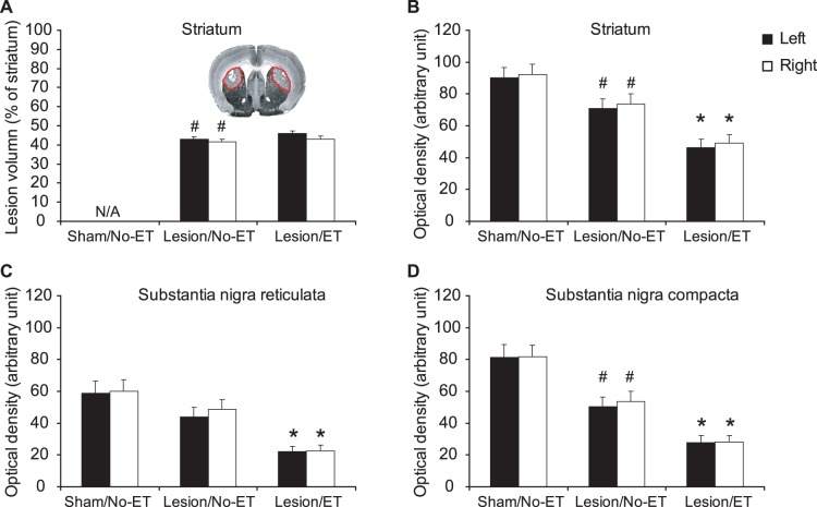
Figure 3. 6-OHDA lesions.
Shown are the effect of bilateral striatal lesions on (A) lesion volume (% of striatal volume), (B) tyrosine hydroxylase (TH) staining of the striatum, (C) TH staining of the substantia nigra reticulata (SNR), and (D) TH staining of the substantia nigra compacta (SNC). #: significant difference between sham rats without exercise training (Sham/No-ET) and lesioned rats without ET (Lesion/No-ET), P<0.05, Student’s t test. *: significant difference between Lesion/No-ET and Lesion/ET, P<0.01, Student’s t test. The inset in (A) shows a representative TH stained coronal section showing bilateral depletion of TH (outlined in red).

