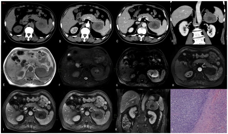
Figure 1. The case of a 66-year-old male patient who had experienced left upper quadrant pain for 1 month is presented.
This patient was diagnosed with PSL at splenectomy. (A) A CT plain scan showed a solitary focal mass with faint hypodensity in the spleen. Contrast-enhanced CT showed mild enhancement of the mass in the (B) arterial phase, (C) the portal phase, and (D) the coronal portal phase. The central hypodensity within the mass represents necrosis. MRI plain scans showed a hypointense signal in (E) T1WI and a hyperintense signal in (F) T2WI and (G) DWI. Enhanced MRI findings for the (H) arterial phase, (I) the portal phase, (J) the delayed phase and (K) the coronal delayed phase demonstrated mild enhancement. (L) The histopathology of the splenic lesion was suggestive of diffuse large B-cell lymphoma (DLBCL) (H&E, 100×).

