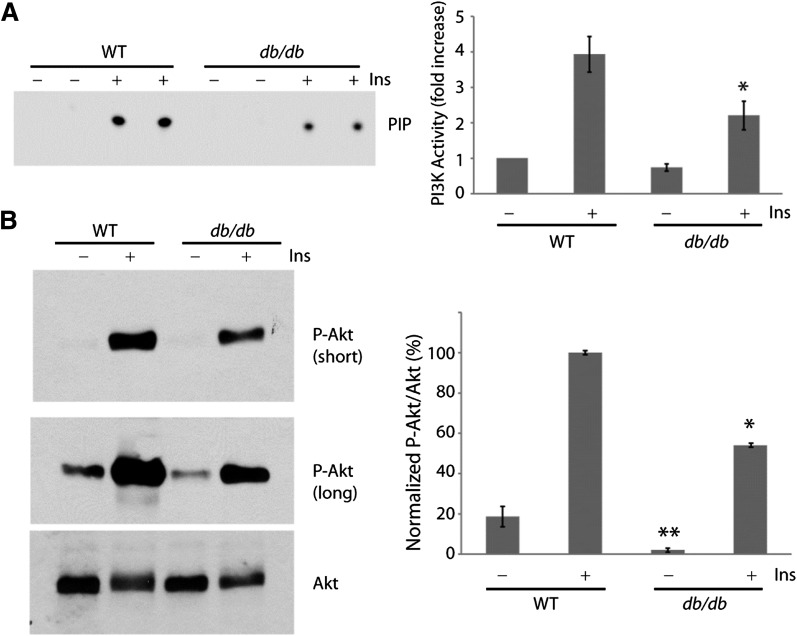
FIG. 4.
Attenuated insulin/PI3K/Akt signaling in db/db hearts. Wild-type (WT) and db/db hearts were perfused with or without 1 unit/L insulin for 10 min while mounted on the Langendorff apparatus. A: Representative autoradiograph of PI3K activity (duplicate measurements) assayed in phosphotyrosine immunoprecipitates of heart lysates (left). Summary graph of normalized PI3K activity (mean ± SE) from three independent experiments (right). *, significantly different from WT treated with insulin (P < 0.05, Student t test). B: Representative Western blots of heart lysates probed sequentially with phospho-Akt (p-Akt) and total Akt antibodies (left). Summary graph of normalized Akt phosphorylation (mean ± SE) from three independent experiments (right). *, significantly different from WT treated with insulin; **, significantly different from untreated WT (P < 0.05, Student t test). Ins, insulin.

