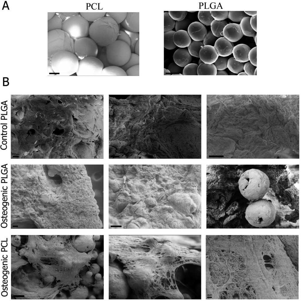
Figure 1.
Representative scanning electron microscopy (SEM) images of week 0 (A) and week 6 (B) samples. Scaffolds were fabricated using PLGA and PCL microspheres at customized processing conditions for sintering (CO2 exposure at 25 bar for 1 h at 25°C followed by depressurization at a rate of 0.101 psi s−1 for PLGA; 45 bar for 4 hrs at 45 °C followed by depressurization at a rate of 0.2 psi s−1 for PCL). Comparable degrees of sintering were observed at week 0, and microsphere shape was better preserved in the PCL constructs by week 6. Chondrogenic PLGA scaffolds are not shown as they did not withstand SEM processing. Scale bars = 100 µm.

