
FIGURE 1.
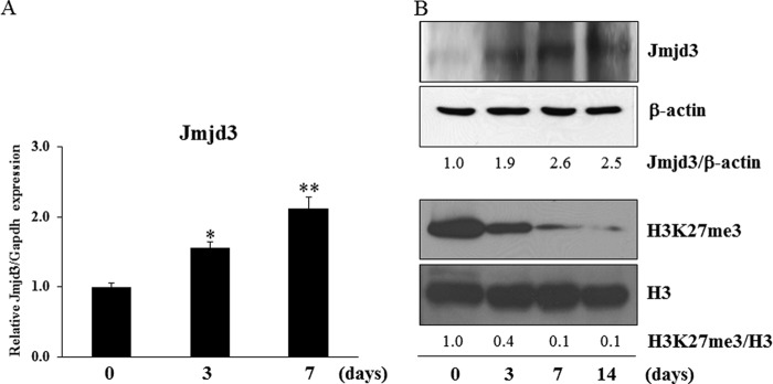
The expression of Jmjd3 in osteoblasts. MC3T3-E1 cells were cultured in the osteoblast differentiation medium for the indicated periods. A, the expression of Jmjd3 was examined by real-time PCR. B, the protein expressions of Jmjd3 and H3K27me3 were examined by Western blot analysis. The relative expressions of Jmjd3/β-actin and H3K27me3/H3 were calculated by computer software (ImageJ). All the results in the real-time PCR are presented as means ± S.E. *, p < 0.05, **, p < 0.01