FIGURE 7.
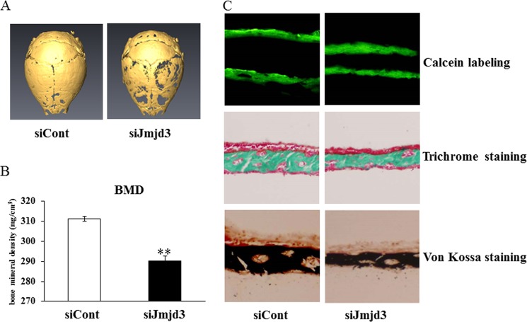
Local administration of siJmjd3 suppressed bone formation in mouse calvaria. A, three-dimensional μCT images of calvaria from mice (12-day-old, n = 4 for siJmjd3 injection, n = 4 for siCont injection) injected with siCont or siJmjd3. B, cortical bone mineral density (BMD) of calvaria was determined by μCT. Data are presented as means ± S.E. **, p < 0.01. C, upper, plastic sections of dissected mouse calvaria were examined under a fluorescence microscope. The distance between two calcein labeling layers reflects the bone mineralization rate. Middle, sections of dissected mice calvaria were stained with Goldner's trichrome to visualize bone thickness. Lower, sections of dissected mice calvaria were stained with von Kossa to visualize bone mineralization.

