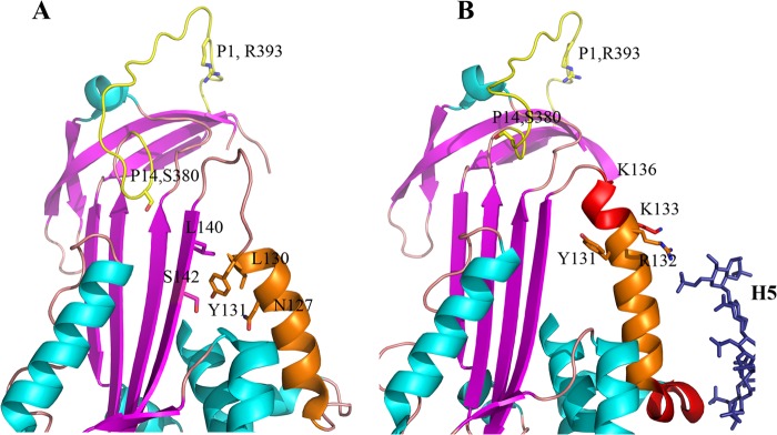
FIGURE 1.

Structural changes in and around helix D of antithrombin upon heparin binding. A, heparin binding region of antithrombin in the heparin-free state (Protein Data Bank code 1E05) (35), showing helix D, the unstructured conformation of residues 131–136 and residues 379 and 380 (P15 and P14) of the reactive center loop inserted into β-sheet A. B, heparin binding region of antithrombin with heparin pentasaccharide bound (Protein Data Bank code 1E03) (35), showing the extension of helix D involving residues 131–136, formation of helix P, and expulsion of residues 379 and 380 of the reactive center loop from β-sheet A.
