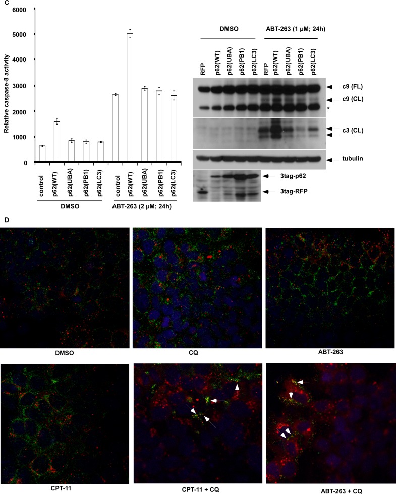
FIGURE 5.
p62/sequestosome 1 co-localizes with caspase-8 aggregates and promotes caspase-8 activation that is disrupted by p62 mutation in ABT-263-treated cells. A, DLD1 cells stably expressing p62 variants (wild-type or mutations in UBA, PB1, and LC3-binding domains) were treated with chloroquine (CQ, 25 μm), and cells were stained with anti-FLAG or anti-LC3 antibodies followed by labeling with fluorescence dye-conjugated secondary antibodies. Red, anti-FLAG; green, anti-LC3; yellow punctae indicate co-localization. The nuclei were counterstained with DAPI (blue). B, HCT116 cells stably expressing a lentiviral mCherry-p62 were transiently transfected with a catalytically inactive procaspase-8 (C360A) fused to Venus N-terminal (VN) or C-terminal (VC). The cells were treated with ABT-263, and localization of mCherry-p62 variants (wild type or mutations in UBA, PB1 domains) and caspase-8-Venus was determined by fluorescence confocal microscopy. Co-localization is shown in merged images (gold color). C, HCT116 cells with stable expression of retroviral p62 variants (wild type or mutations in UBA, PB1 and LC3-binding domains) were treated with ABT-263, and activation of caspase-8 (left) and cleavage of caspase-9 and -3 (right) were determined. Red fluorescence protein (RFP) serves as a control. *, nonspecific band. D, DLD1 cells were treated with ABT-263 (2 μm) or CPT-11 (10 μm) alone or combined with chloroquine (25 μm) for 12 h, and immunofluorescence staining of p62 and caspase-8 was performed. Red, p62; green, caspase-8; nuclei are counterstained with DAPI (blue). Co-localization of caspase-8 with p62 is shown by yellow punctae. DMSO, dimethyl sulfoxide.


