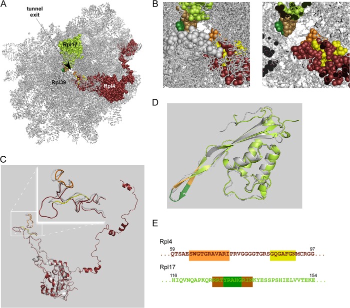
FIGURE 1.
The ribosomal tunnel proteins of yeast. A, view of the Saccharomyces cerevisiae large ribosomal subunit based on PDB code 3U5H/3U5I (7, 10). Shown is the ribosomal RNA (gray) of the large subunit and the three tunnel proteins Rpl4 (purple), Rpl17 (lime), and Rpl39 (gray). B, enlarged image section of the tunnel exit region. Left, exterior view; right, same image section but with a clipping plane at the constriction site of the tunnel. Rpl4 (purple, flexible residues in orange, insertion segment in yellow); Rpl17 (lime, flexible tip of the β-hairpin in green, hinges in brown). C, superposition of S. cerevisiae Rpl4 (colors are as described in A, PDB code 3U5I) and Haloarcula marismortui L4 (gray, PDB code 2QA4). The inset shows a close-up of the tunnel exposed segments of Rpl4/L4. The flexible segment of S. cerevisiae Rpl4 (orange) is structurally similar to the corresponding segment of H. marismortui L4. The insertion segment (yellow) leads to the formation of an extended loop in Rpl4. D, superposition of S. cerevisiae Rpl17 (color code as described in A, PDB code 3U5I) and H. marismortui L22 (gray, PDB code 2QA4) reveals structural similarity. E, partial amino acid sequences of S. cerevisiae Rpl4 (amino acids 59–97) and S. cerevisiae Rpl17 (amino acids 116–154), containing the tunnel exposed flexible segments. Rpl4, orange, flexible residues; yellow, insertion segment; Rpl17, green, flexible tip, brown, hinges.

