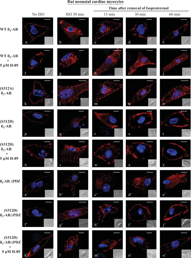
FIGURE 3.
Characterization of β1-AR trafficking in rat neonatal ventricular myocytes. Neonatal rat cardiac myocytes were cultured on collagen-coated coverslips and then infected with 100 multiplicities of infection of adenovirus harboring FLAG-tagged WT β1-AR or the other β1-AR constructs. Cells were prelabeled for 1 h with Cy3-anti-FLAG antibody and fixed (a, f, k, p, u, a′, f′, and k′). The rest of the slides were exposed to isoproterenol (ISO) for 30 min then fixed (b, g, l, q, v, b′, g′, and l′). The remaining slides were subjected to recycling conditions for the indicated time period and then fixed and visualized by confocal microscopy using the Olympus FluoViewTM FV-1000 confocal microscope (n = 3 experiments and at least 40 images per experiment were analyzed). Each scale bar, 5 μm.

