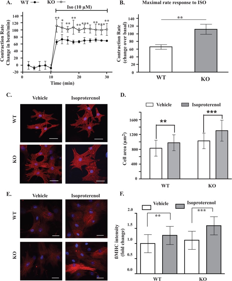
FIGURE 6.
Effect of AKAP5 expression on cardiomyocyte function. A, contraction rates of cardiac myocytes prepared from AKAP5+/+ or AKAP5−/− mice were measured for 10 min before and for 30 min after the addition of 10 μm isoproterenol (ISO). B, increases in maximal contraction rates in A were compared between AKAP5+/+ myocytes and AKAP5−/− myocytes. C and E, cardiomyocytes from AKAP5+/+ mice or AKAP5−/− mice were stimulated with vehicle or 10 μm isoproterenol for 6 h and then fixed and stained for α-actinin (red) and β-myosin heavy chains (red). D and F, 80 cells per group per isolation were analyzed by morphometry in D and for fluorescence intensity in F. Each scale bar, 20 μm. Bars represent means ± S.E. A, analysis of variance was determined by Student's t test. **, p < 0.01, and ***, p < 0.001. B and D, **, p < 0.01, and ***, p < 0.001 for isoproterenol versus basal morphometry in WT and KO cells, respectively. **, p < 0.01, and ***, p < 0.001 for isoproterenol versus basal β-MHC in WT and KO respectively; one-way ANOVA with Newman-Keuls post tests.

