
FIGURE 4.
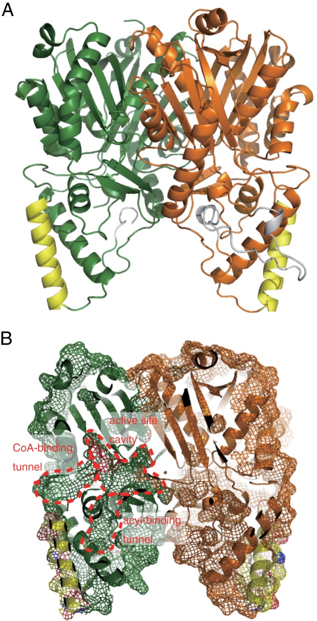
Overall structure of ArsC. A, ribbon drawing diagram of the ArsC dimer. Each monomer is shown in green and orange. Helix α3 and the flexible insertional loop region are shown in yellow and gray, respectively. B, molecular surface of the ArsC dimer focusing on the inside of the enzyme. The CoA binding tunnel, active site cavity, and acyl binding tunnel are indicated.