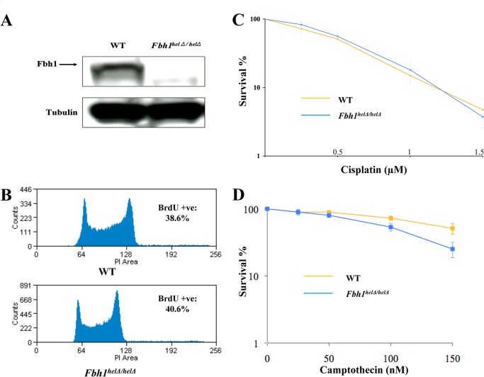
FIGURE 1.
Characterization of Fbh1helΔ/helΔ cells. A, Fbh1helΔ/helΔ cells do not express any full-length FBH1 protein. Western blot of an extract from WT and Fbh1helΔ/helΔ cells was immunoblotted for FBH1 and tubulin as a loading control. B, cell cycle distribution is only marginally altered in Fbh1helΔ/helΔ cells compared with WT cells by FACS analysis. C, clonogenic survival of WT and Fbh1helΔ/helΔ cells following exposure to cisplatin. D, clonogenic survival of WT and Fbh1helΔ/helΔ cells following exposure to camptothecin. The means and the S.E. (error bars) are shown for three independent experiments.

