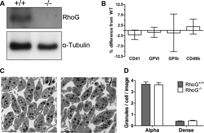
FIGURE 2.
A, immunoblotting confirmed RhoG protein expression in RhoG+/+ mouse platelets and ablation of RhoG protein expression in platelets from constitutive RhoG knock-out mice (RhoG−/−). B, flow cytometry was used to evaluate the expression of key platelet surface glycoproteins in RhoG−/− mice. For each experiment, surface expression was determined in duplicate by evaluating the binding of FITC-conjugated monoclonal antibodies. Isotype antibody controls were used to account for nonspecific binding. There were no significant differences between WT and RhoG−/− platelet expression of CD41, GPVI, GPIb, or CD49b. Bars represent the mean, and error bars represent S.E. (n = 10). C, to evaluate RhoG−/− platelet ultrastructure, ultrathin sections were imaged with a transmission electron microscope. Subjectively, there was no difference in the subcellular morphology of WT platelets compared with those lacking RhoG. Scale bars = 2 μm. D, the numbers of platelet α-granules and dense granules were evaluated using transmission electron microscopic sections taken at ×2800 magnification. The total numbers of platelet granules in 10 equivalent-sized fields of view were quantified manually using ImageJ, and granule numbers are expressed as granules/cell/image. Bars represent means ± S.E. of three mice/group. There was no significant difference in the numbers of α-granules or dense granules between WT and RhoG−/− platelets.

