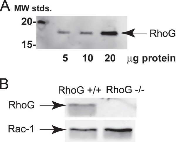
FIGURE 1.

Expression of RhoG in human and mouse platelets. A, increasing amounts of human platelet lysate (in μg) were separated by SDS-PAGE, Western-blotted, and probed with anti-RhoG antibody. MW stds., molecular weight standards. B, RhoG expression in platelet lysates from RhoG-null mice (RhoG−/−) and wild-type littermates (RhoG+/+) was probed by Western blotting RhoG. Rac1 was probed to verify protein loading. The blot shown is representative of three independent experiments.
