FIGURE 7.
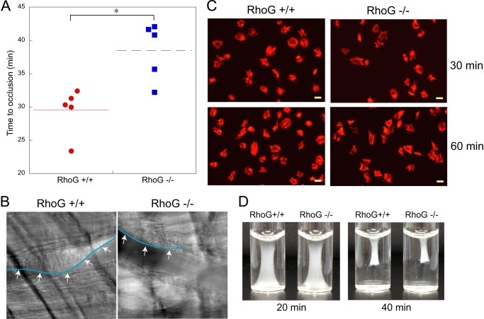
Analysis of thrombus formation, platelet spreading, and clot retraction in RhoG−/− mice. A, the time required for occlusion of cremaster arterioles in RhoG+/+ and RhoG−/− mice was measured using the microvascular thrombosis model with light/dye-induced injury as described under “Experimental Procedures.” Five mice of each genotype were used, and statistical analysis revealed a significant difference between the two genotypes of mice. *, p < 0.01. B, representative images of cremaster arterioles were taken from RhoG+/+ and RhoG−/− mice 30 min after injury. As seen by the outline (arrows) of the thrombus formed, thrombus formation was inhibited in RhoG−/− mice. C, washed platelets (1 × 10⁷ platelets/ml) from RhoG+/+ and RhoG−/− mice were plated on fibrinogen-coated coverslips. After washing three times with PBS, adherent cells were fixed with 3.7% paraformaldehyde, stained with rhodamine-phalloidin, and analyzed by fluorescence microscopy. D, clot retraction analysis of platelets from RhoG+/+ and RhoG−/− mice. Images are representative of three independent experiments.

