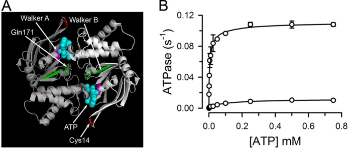
FIGURE 1.

Structure and function of the NBD dimer. A, structure of the NBD dimer. Each monomer is represented in a different tone of gray. Walker motifs A and B are labeled and shown in purple and green, respectively. The two ATPs (cyan, spheres), Cys-14 (red, sticks), and Gln-171 (green, sticks) are also labeled. The figure displays the structure of the nucleotide-bound MJI based on PDB 1L2T. Glu replaces Gln-171 in MJ. B, dependence of the ATPase activity on ATP concentration. Data are means ± S.E. of two independent measurements. The line is a fit of the Hill equation to the data.
