
Figure 1.
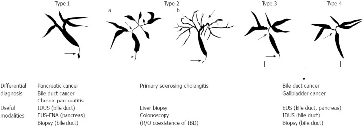
Cholangiographic classification of IgG4-related sclerosing cholangitis and differential diagnosis. Stenosis is located only in the lower part of the common bile duct in type 1; stenosis is diffusely distributed in the intra-and extra-hepatic bile ducts in type 2. Type 2 is further subdivided into two. Extended narrowing of the intrahepatic bile ducts with prestenotic dilation is widely distributed in type 2a. Narrowing of the intrahepatic bile ducts without prestenotic dilation and reduced bile duct branches are widely distributed in type 2b; stenosis is detected in both the hilar hepatic lesions and the lower part of the common bile ducts in type 3; strictures of the bile duct are detected only in the hilar hepatic lesions in type 4. IDUS: Intraductal ultrasonography; EUS-FNA: Endoscopic ultrasound-guided fine needle aspiration; IBD: Inflammatory bowel disease.