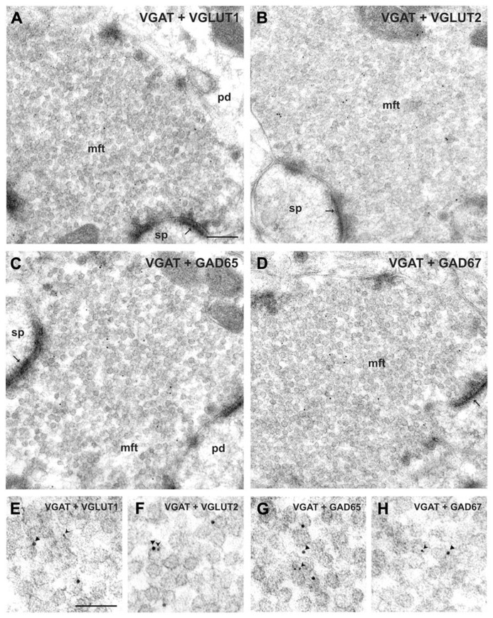
FIGURE 1.
Co-localization of VGAT with either GAD or VGLUT isoforms in hippocampal mossy fiber terminals. (A,B) Mossy fiber terminals (mft) of the CA3 area were double-immunolabeled with the guinea pig antiserum against VGAT (10 nm gold particles, arrowheads in E, F) and rabbit antisera against either VGLUT1 and VGLUT2 (5 nm gold particles, forked arrowheads in E, F, respectively). Note the SV exhibiting putatively co-localized immunogold signals for VGAT and VGLUT2 shown in (F). (C,D) Mossy fiber terminals were double-immunolabeled with the guinea pig antiserum against VGAT (10 nm gold particles, arrowheads in G, H) and rabbit antisera against either GAD65 or GAD67 (5 nm gold particles each, forked arrowheads in G, H), respectively. (E–H) Represents details at higher magnification. pd, pyramidal dendrite; sp, spine; thin arrows indicate asymmetric contacts. Bars given represent: (A–D), 200 nm; (E–H), 100 nm (from Zander et al., 2010).

