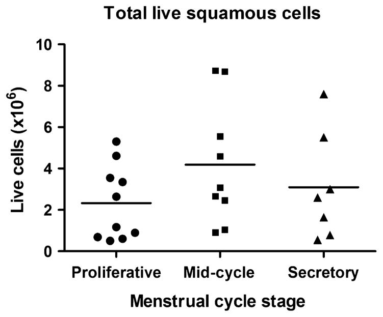
Fig. 2.
Recovery of vaginal epithelial cells. Cells were recovered from menstrual cups obtained during three stages of the menstrual cycle: proliferative (d6–8), mid-cycle (d13–15) and secretory (d21–23). Viability was determined by trypan blue exclusion. Each dot represents the number of live cells in individual menstrual cups.

