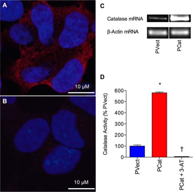
Figure 1.

Characterization of catalase gene overexpression in SH-SY5Y neuronal cells. (A) Immunocytochemistry of human SH-SY5Y neuronal cells stably expressing the catalase gene vector (PCat) showing localization of catalase in the cytoplasm. (B) Immunocytochemistry of human SH-SY5Y neuronal cells stably expressing the pcDNA4/TO/myc–His expression vector (PVect) showing low level localization of catalase above background. Catalase appears red (CAT 505 monoclonal anti-catalase staining), and the nucleus appears blue (TO-PRO-3 iodide staining). Bars = 10 μm. (C) RT-PCR analysis of catalase and β-actin mRNA in human SH-SY5Y neuronal cells stably expressing the catalase gene vector (PCat) and stably expressing the pcDNA4/TO/myc–His expression vector (PVect). (D) Catalase activity in extracts from Human SH-SY5Y neuronal cells stably expressing the catalase gene vector (PCat) and stably expressing the pcDNA4/TO/myc–His expression vector (PVect), plus the effect of the catalase inhibitor 50 mM 3-AT on PCat cell extract catalase activity. Results are expressed as % PVect catalase activity (mean ± SEM). *P < 0.05 vs PVect extracts; †P < 0.05 vs PCat extracts; one-way ANOVA.
