
Figure 8.
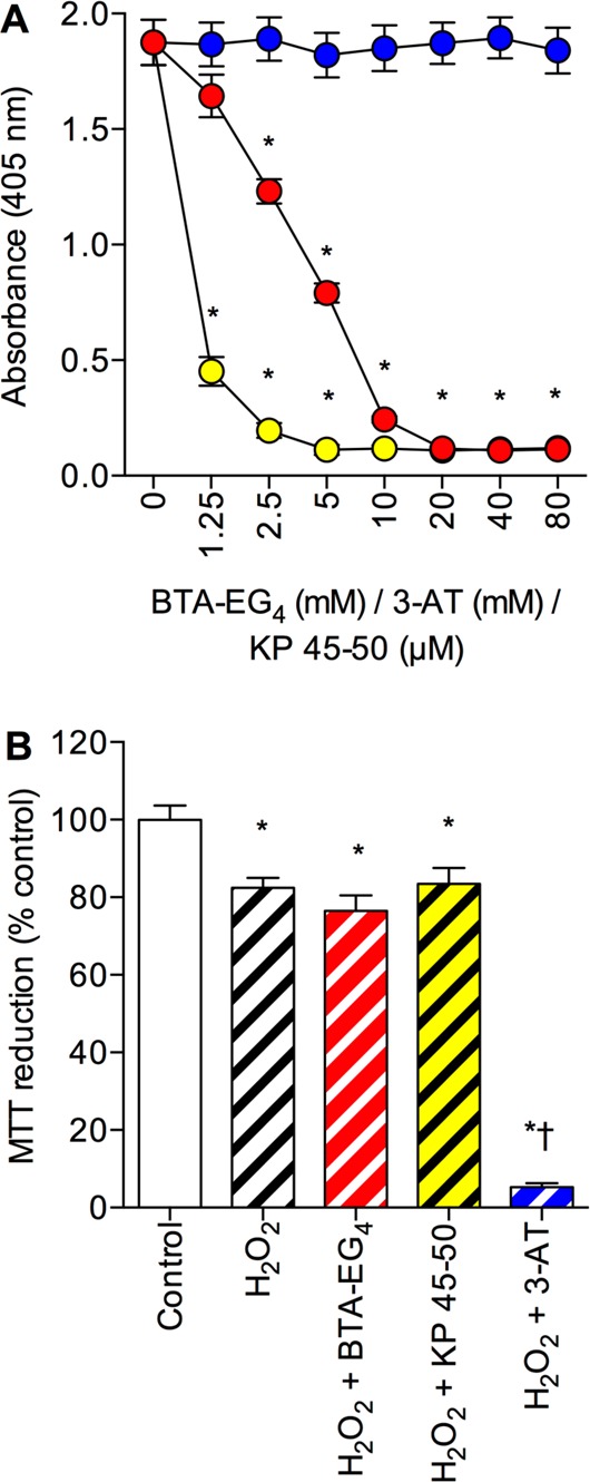
Effects of BTA-EG4, KP 45–50, and 3-AT on amyloid-β binding to catalase and protection against H2O2 toxicity. (A) Catalase coated plates were incubated with 1 μM biotinylated Aβ 1–42 in the presence of 0–80 mM 3-AT (closed blue circles), 0–80 mM BTA-EG4 (closed red circles), or 0–80 μM KP 45–50 (closed yellow circles) and bound material determined by EIA. *P < 0.05 vs biotinyl-Aβ 1–42 alone; one-way ANOVA. (B) Human SH-SY5Y neuronal cells stably expressing the catalase gene vector (PCat) were pretreated with medium alone (clear hatched column), 20 μM BTA-EG4 (red hatched column), 20 μM KP 45–50 (yellow hatched column), or 50 mM 3-AT (blue hatched column) prior to exposure to 500 μM H2O2 and cell viability determined by MTT reduction. Control was medium alone (clear column). Results are mean ± SEM; *P < 0.05 vs control; †P < 0.05 vs H2O2 alone; one-way ANOVA.