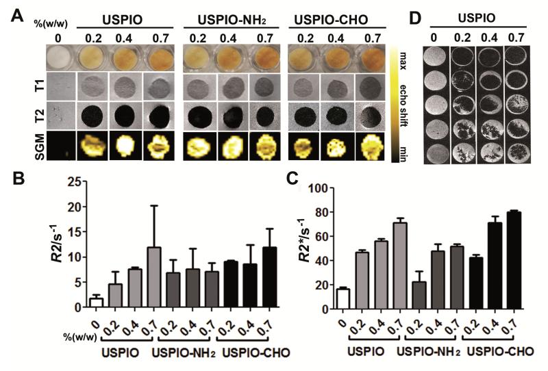
Figure 3. MR imaging of collagen scaffolds labeled both passively and actively with USPIO.
A) Visual depiction, T1- and T2-eighted MRI, and Susceptibility Gradient Mapping (SGM) of labeled scaffolds. B-C) Quantitative R2- (B) and R2*-relaxometry (C) analysis. All labeled scaffolds were clearly detectable in MRI and showed significant increases in R2 and R2* relaxation rates with increasing amounts of incorporated USPIO. D) MR monitoring of scaffold degradation over time upon exposure to collagenase.

