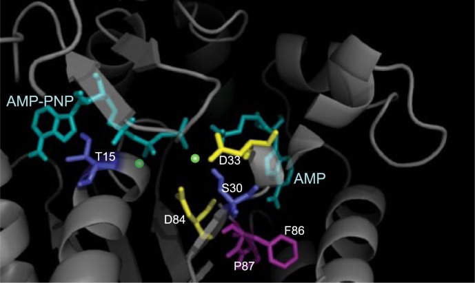
FIGURE 6.
A close-up crystal structure of E. coli AK ligand binding sites (1ANK (61)). The substrates AMP-PNP and AMP are drawn in cyan. Residues Asp-33 and Asp-84 (yellow) as well as residues Ser-33 and Thr-15 (deep blue) are potential magnesium binding residues. The postulated magnesium ion binding locations, (green dots) are in between these residues and the β- and γ-phosphate groups of AMP-PNP. Residues Phe-86 and Pro-87 (magenta) have been shown to interfere with the “AMP inhibition behavior” (35, 36) and are located near the AMP binding site.

