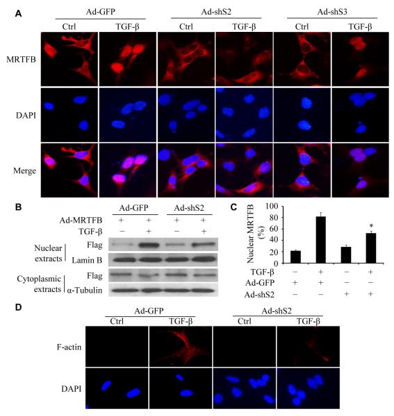
Figure 6. Smad2 was essential for MRTFB nuclear translocation.
(A) Knockdown of Smad2 blocked MRTFB nuclear translocation. Monc-1 cells were transduced with adenovirus expressing Flag-tagged MRTFB and GFP (Ad-GFP), Smad2 (Ad-shS2) or Smad3 shRNA (Ad-shS3) as indicated for 2 days followed by vehicle (Ctrl) or TGF-β induction for 2 hours. MRTFB expression and its cellular location were detected by immunostaining with anti-Flag antibody. DAPI stains nuclei. TGF-β induced MRTFB nuclear translocation, which was blocked by Ad-ShS2, but not Ad-shS3. (B) MRTFB cellular location detected by western blot. Monc-1 cells were treated the same as in A, and cytoplasmic or nuclear MRTFB was detected with Flag antibody. (C) Nuclear MRTFB in B was quantified and shown as percentage of the total MRTFB. Prior to the calculation, nuclear MRTFB was normalized to Lamin B, and cytoplasmic MRTFB was normalized to α-Tubulin. *P<0.05 compared to Ad-GFP-transduced cells treated with TGF-β. (D) Knockdown of Smad2 blocked F-actin formation. Monc-1 cells were transduced with Ad-GFP or Ad-ShS2 followed by vehicle (Ctrl) or TGF-β treatment. F-actin was stained with Phalloidin.

