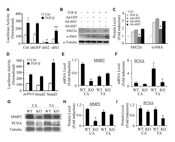
Figure 8. Progenitor-specific roles of Smad2 and Smad3 in regulating VSMC marker gene transcription and protein expression.
(A) Smad2 was more important than Smad3 in regulating VSMC marker gene promoter activity in NCCs. Monc-1 cells were transduced with adenovirus expressing GFP (shGFP), Smad2 (shS2), or Smad3 (shS3) shRNA for 2 days and transfected with SM22α promoter followed by vehicle (Ctrl) or TGF-β (5 ng/ml) treatment. Luciferase assays were performed. Luciferase activity was normalized to renilla activity. *P<0.001, **P<0.05 compared to shGFP/TGF-treated groups. (B) Smad2, but not Smad3, was essential for VSMC mark protein expression in NCCs. Monc-1 cells were treated similarly as described in A without promoter transfection. α-SMA and SM22α protein expression was detected by western blot. (C) Quantification of protein expression shown in B by normalized to α-tubulin. *P<0.01 compared to Ad-GFP group for α-SMA and SM22α, respectively. (D) Smad3 was more important than Smad2 in VSMC differentiation from mesenchymal progenitors. 10T1/2 cells were co-transfected with SM22α promoter and pcDNA, Smad2, or Smad3 cDNA followed by vehicle (Ctrl) or TGF-β treatment. Luciferase assays were performed. Luciferase activity was normalized to renilla activity. *P<0.01 compared to pcDNA/TGF-β-treated group. (E and F) Smad2 was deleted in SMCs by crossing Smad2floxed mice with SM22α-Cre mice. Carotid (CA) or thoracic artery (TA) media layers from wild type littermates (WT) or SMC-specific Smad2 knockout mice (KO) were homogenized, and total RNA was extracted. qPCR were performed to detect MMP2 (E) and PCNA expression (F) and fold inductions were shown. *P<0.01 compared to the corresponding WT SMCs. (G) MMP2 and PCNA protein expression in WT and Smad2 KO SMCs was examined by western blot. (H-I) MMP2 (H) and PCNA expression (I) was normalized to α-tubulin. *P<0.01 compared to the corresponding WT SMCs.

