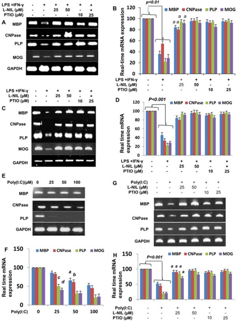
Figure 3. Effect of L-NIL and PTIO on (IFN-γ + LPS)- and polyIC-mediated decrease in myelin gene expression in human fetal mixed glial cultures.
Human primary brain (A & B) and spinal cord (C & D) mixed glial cells preincubated with different concentrations of L-NIL and PTIO for 1 h were stimulated with 1 µg/ml of LPS and 6.25 mU/ml of IFN-γ. After 24 h of stimulation, the expression of MBP, MOG, PLP, and CNPase mRNA was analyzed in cells by semi-quantitative RT-PCR (A & C). Quantitative real time PCR was also employed to further clarify the expression of myelin genes (B & D). ap<0.001. Human primary mixed glial cell were stimulated with different concentrations of polyIC under serum free conditions. After 24 h of stimulation, the expression of myelin genes was analyzed in cells by semi-quantitative RT-PCR (E) and real time-PCR (F). ap<0.01, bp<0.01, cp<0.001 & dp<0.001 vs. polyIC-treated cells. Cells preincubated with different concentrations of L-NIL and PTIO for 1 h were stimulated with 100 µg/ml of polyIC. After 24 h of stimulation, the expression of MBP, MOG, PLP, and CNPase mRNA was analyzed in cells by semi-quantitative RT-PCR (G) and Quantitative real time PCR (H). Results are mean ± S.D. of three different experiments. ap<0.001 vs. polyIC-treated cells.

