
Figure 2.
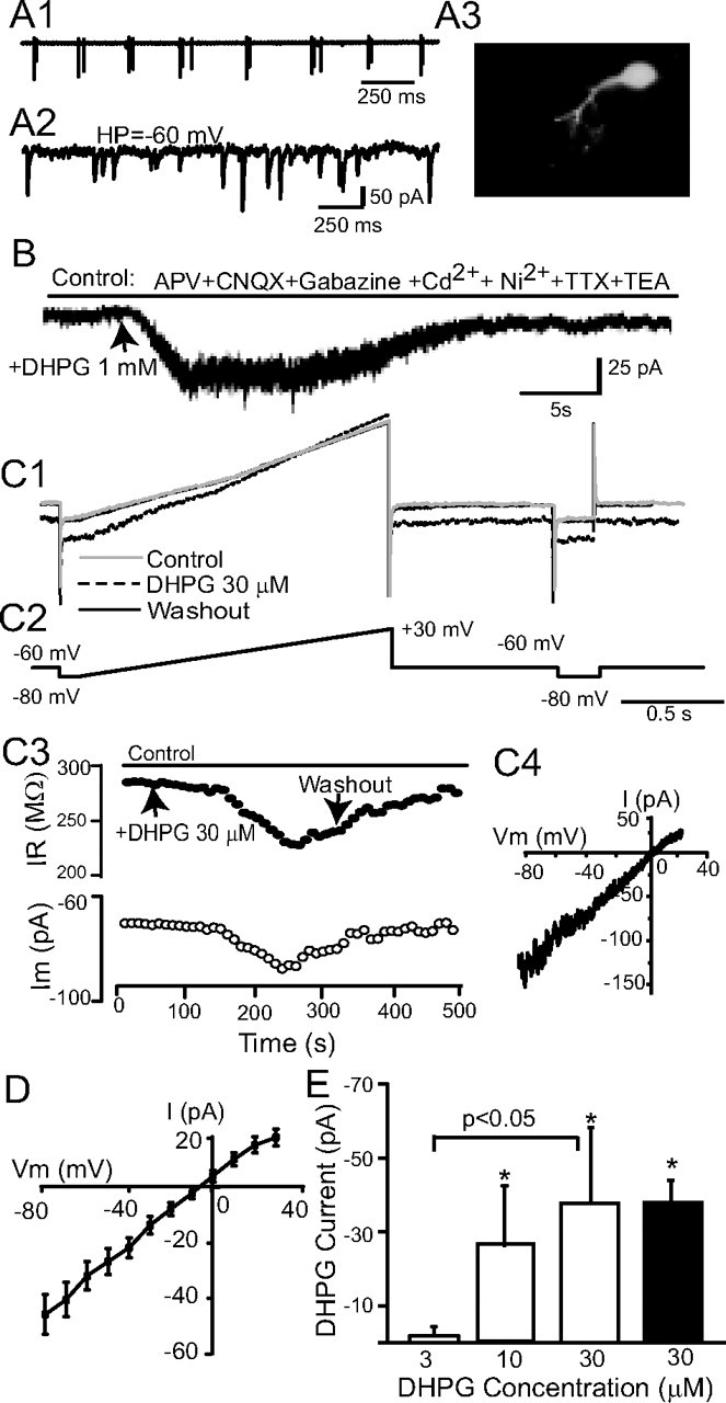
DHPG evokes an inward current in ET cells. A, An ET cell recorded in whole-cell voltage-clamp mode was initially identified on the basis of extracellularly recorded spike bursts (A1). In voltage-clamp mode, sEPSCs occurred randomly (A2). A3, Lucifer yellow staining shows the morphology of this typical ET cell. B, A focal puff of DHPG (1 mm) adjacent to this ET cell evoked an inward current at a HP of −60 mV. C1, C2, Membrane currents (C1) produced by slow voltage ramps (C2, command voltages) were used to generate I–V curves before and during bath-applied DHPG (30 μm), and again after washout; at right, hyperpolarizing pulses monitored changes in input resistance. C3, The inward current elicited by bath-applied DHPG is accompanied by decreased membrane resistance (HP = −60 mV). C4, The DHPG-evoked current for a single ET cell, obtained by subtraction of the control and DHPG I–V curves in C1, was linear and reversed polarity near 0 mV. D, Mean I–V plot of the DHPG current in 27 ET cells; the mean reversal potential was −5.2 ± 2.3 mV. E, Dose–response relationship of DHPG-evoked currents in ET cells. The magnitude of the inward current elicited by cumulative doses of 3, 10, and 30 μm DHPG were measured in individual ET cells (open bars, n = 6, HP = −60 mV); 10 and 30 μm DHPG significantly increased the amplitude of the inward current relative to baseline (*p < 0.05 one-way repeated-measures ANOVA followed by Newman–Keuls tests), and the currents evoked by 30 μm were significantly greater than that evoked by 3 μm (p < 0.05, Newman–Keuls test). The amplitude of the inward current elicited by a single 30 μm application of DHPG (black bar, n = 27) did not differ from that elicited in the cumulative condition (p > 0.05, unpaired t test).