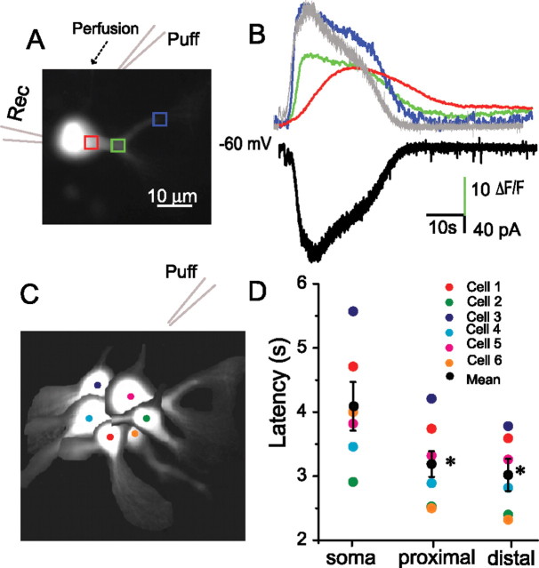
Figure 5.
DHPG-evoked [Ca2+]i transients originate in ET cell dendrites. A, Fluorescence image of an ET cell recorded with a pipette containing 100 μm fura-2 and voltage clamped at −60 mV. B, Time course of changes in [Ca2+]i in the soma (red trace), proximal (green trace) and distal (blue trace) dendritic regions (corresponding to the rectangles in A1), and inward current (black trace) elicited by a focal puff of DHPG (1 mm) 30 μm upstream from the soma; note that the time course of the inward current (inverted trace shown in gray) closely corresponds to that of the Ca2+ transient in the distal dendrite. Experiment was in conducted in normal ACSF (no antagonists). C, Schematic diagram showing the position of six sampled ET cells with respect to the puffer pipette. D, Individual cell and group data (mean ± SEM, black circles) showing that the latencies of [Ca2+]i transients in the soma were longer than those in the dendrites (*p < 0.001, n = 6).

